After two decades in medical technology, I have watched robotic systems evolve from novelty to necessity. Today, artificial intelligence is not replacing surgeons; it is giving them superpowers. But understanding what AI actually does in the OR is critical before you consider adopting these tools. Let me cut through the hype.
What AI brings to the table is real-time data fusion. The most advanced systems combine preoperative imaging, such as CT or MRI, with live video from the endoscope. The AI overlays critical structures like blood vessels, ureters, or tumor margins directly onto the surgical field. This is not a gimmick. In my experience, it reduces cognitive load significantly. You no longer have to mentally register a 2D scan with a 3D anatomy. The machine does that for you.
The key features you should evaluate are threefold. First, segmentation accuracy. How well does the AI identify and highlight structures? Look for systems that have been trained on thousands of cases, not just lab models. Second, latency. The overlay must be near-instant, under 100 milliseconds. Any delay creates a dangerous disconnect between what you see and what the robot does. Third, adaptability. Does the AI adjust to your specific patient anatomy? Some systems allow you to manually correct the overlay if the AI misidentifies a structure. This is essential.
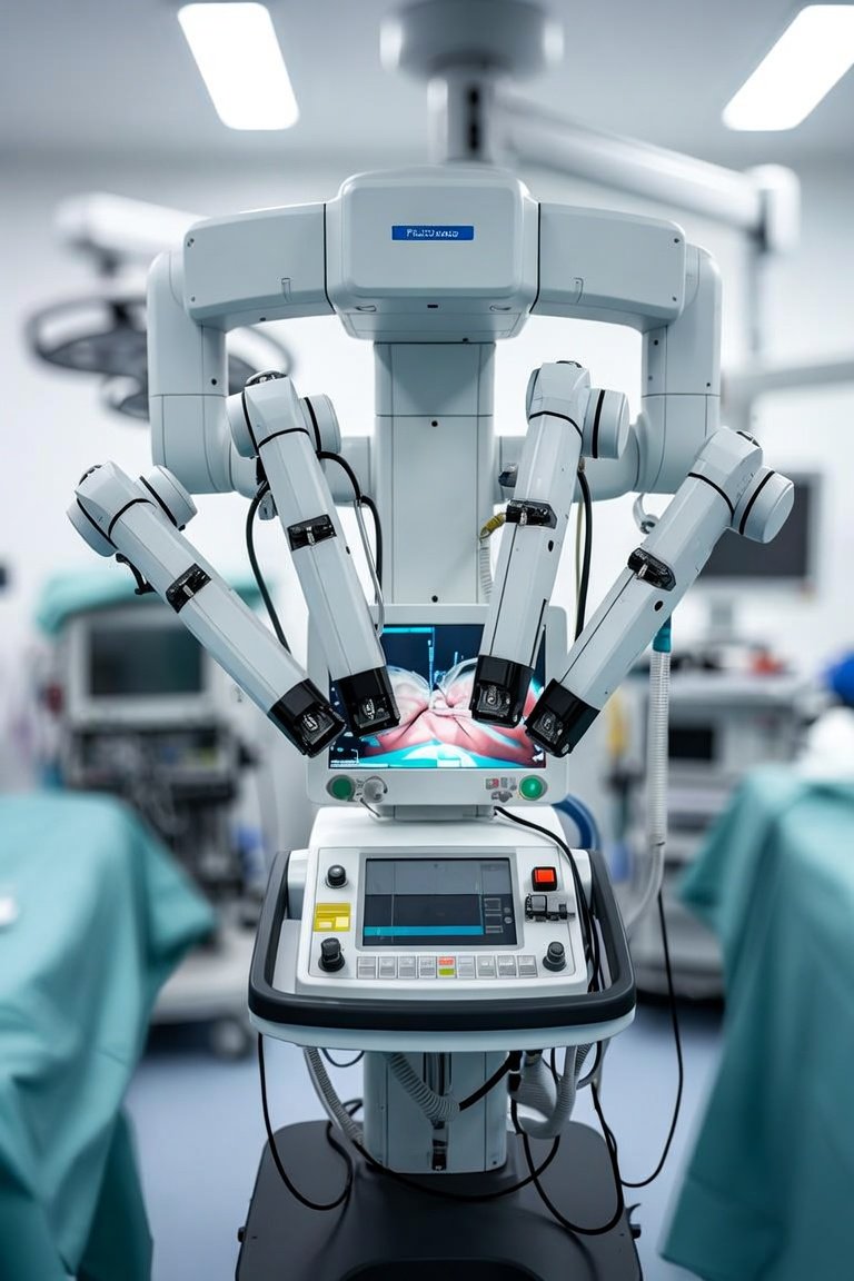
When comparing platforms, you have two main categories. The first is integrated systems, where AI is built into the robotic console. Examples include the da Vinci SP with Firefly fluorescence and newer platforms from companies like Medtronic and Johnson & Johnson. These offer seamless workflow but come with a high capital cost. The second category is AI modules that attach to existing laparoscopic towers. These are more affordable and can be upgraded without replacing your entire system. I have seen excellent results with both, but the choice depends on your volume and budget.
What should you look for when evaluating a system? Start with the training data. Ask the vendor how many surgical cases were used to train the AI. More is better, but diversity matters too. A system trained only on bariatric cases may fail in colorectal surgery. Next, test the system yourself. Run it on recorded cases from your own practice. See how it handles challenging anatomy like scar tissue or variant vessels. Finally, demand a clear upgrade path. AI improves rapidly. You need a system that can receive software updates without hardware replacement.
In the real world, AI-assisted surgery shines in two areas. First, in complex oncologic resections where margin status is critical. The AI can highlight tumor boundaries that are invisible to the naked eye. Second, in procedures with high risk of vascular injury, like pancreatic or liver surgery. The overlay shows vessels that might be hidden behind fat or adhesions. I have personally seen surgeons reduce blood loss by 30 percent using these systems.
My recommendation is to start small. Do not try to adopt AI for every case immediately. Pick one high-volume, moderately complex procedure where the technology can prove itself. Laparoscopic cholecystectomy or partial nephrectomy are excellent starting points. Train your team thoroughly, including the nursing and anesthesia staff. The machine is only as good as the people running it.
AI-assisted surgery is here to stay. It will not make you obsolete. It will make you safer, faster, and more precise. But only if you understand what it can and cannot do. Invest the time to learn it properly. Your patients will thank you.
Innovaciones y Tecnología
AI-Assisted Surgery: A Practical Guide for the Operating Room