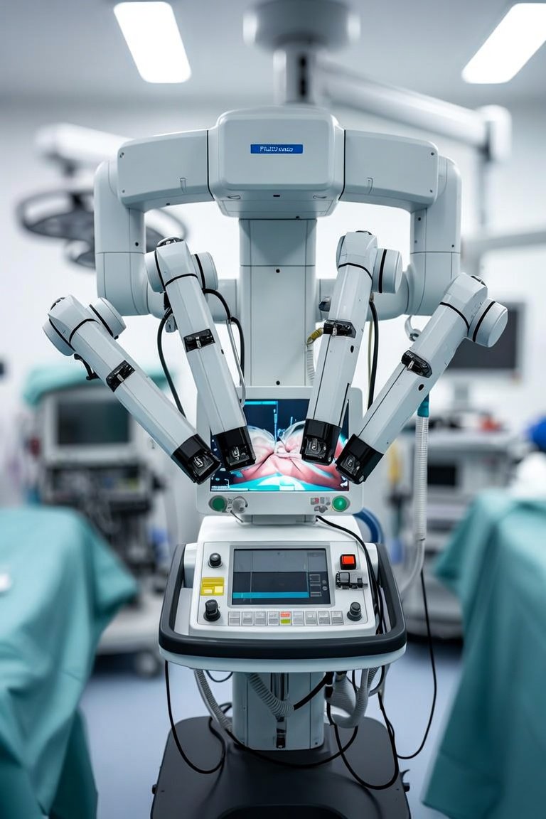
You have likely seen the headlines, but the real question is how AI-assisted surgery changes your daily workflow in the operating room. After two decades of working alongside surgeons and evaluating hundreds of systems, I can tell you this is not science fiction. It is a practical evolution of existing robotic platforms and imaging systems that adds a layer of intelligent decision support. The core idea is simple: AI algorithms analyze real-time data from cameras, sensors, and preoperative scans to give you enhanced visualization, instrument guidance, and even predictive warnings. This is not about replacing your hands. It is about extending your perception.
The first thing to understand is the three main categories of AI assistance you will encounter in the OR. Number one is augmented reality overlays. Here, AI fuses live endoscopic video with preoperative CT or MRI data, highlighting critical structures like blood vessels, ureters, or tumor margins directly on your screen. This reduces the mental load of mentally mapping 2D images onto a 3D field. Number two is instrument tracking and guidance. The AI can recognize your current tool, its tip position, and its trajectory. It can then issue a visual or audible alert if you approach a no-go zone, such as the optic nerve during sinus surgery or a major vessel during laparoscopy. Number three is tissue characterization. Advanced systems use hyperspectral imaging or optical coherence tomography combined with AI to differentiate healthy tissue from cancerous tissue in real time, giving you a biopsy-level assessment without waiting for pathology.
When comparing systems, focus on the integration depth. Some platforms are add-on software for existing robotic arms, like the da Vinci SP with its Firefly fluorescence and AI-enhanced vision. Others are standalone modules that work with standard laparoscopes. The key differentiator is the AI model's training data. Ask your vendor specifically: was the algorithm trained on data from your specialty, your typical patient demographics, and your common procedures? A model trained on general abdominal surgery will not perform well for urologic or thoracic cases. Also, check the latency. The AI must process and overlay information in under 200 milliseconds to be useful during active dissection. Anything slower becomes a distraction.
What should you look for when evaluating a system for your hospital? First, demand a hands-on simulation session with your own team. The AI should not require a separate technician to operate. It must integrate into your existing foot pedal or voice commands. Second, verify the regulatory clearance. In the United States, look for FDA 510(k) clearance specifically for the AI feature, not just the base device. Third, examine the alarm fatigue risk. A good system has adjustable sensitivity thresholds so you are not bombarded with false positives during routine dissection. Finally, consider the data privacy. The AI will record video and metadata. Ensure your hospital has a clear policy on how that data is stored, anonymized, and used for future model updates.
My recommendation is to start with a focused rollout. Pick one high-volume, high-risk procedure in your specialty, such as laparoscopic cholecystectomy or robotic prostatectomy. Use the AI overlay for one month, track your complication rates, operative time, and subjective confidence. Compare that against your historical data. The technology is powerful, but it demands validation in your hands. AI-assisted surgery is here to stay. The doctors who embrace it with a critical, informed eye will be the ones who see the clearest benefit for their patients.
Innovaciones y Tecnología
AI-Assisted Surgery: What Doctors Need to Know