Over the past two decades, I have watched surgical robotics evolve from experimental curiosities into tools that now influence nearly every operating room. But the term AI-assisted surgery is often misunderstood. Let me clarify what it actually means for you, the practicing surgeon, and how to evaluate these systems without getting lost in marketing.
First, understand that current AI in surgery is not autonomous. It is a decision-support layer. The core features you will encounter are threefold. 1) Preoperative planning: AI algorithms analyze CT or MRI scans to create 3D models, highlight critical structures like vessels or nerves, and simulate access routes. This is particularly useful in complex spine, liver, or lung resections. 2) Intraoperative guidance: During surgery, AI overlays real-time data onto the endoscopic or robotic view. For example, it can track instrument tips, measure tissue perfusion using near-infrared fluorescence, or alert you if you approach a known danger zone. 3) Postoperative analysis: Some platforms now record surgical video and automatically tag key steps. This is invaluable for training, auditing, and identifying patterns that lead to complications.
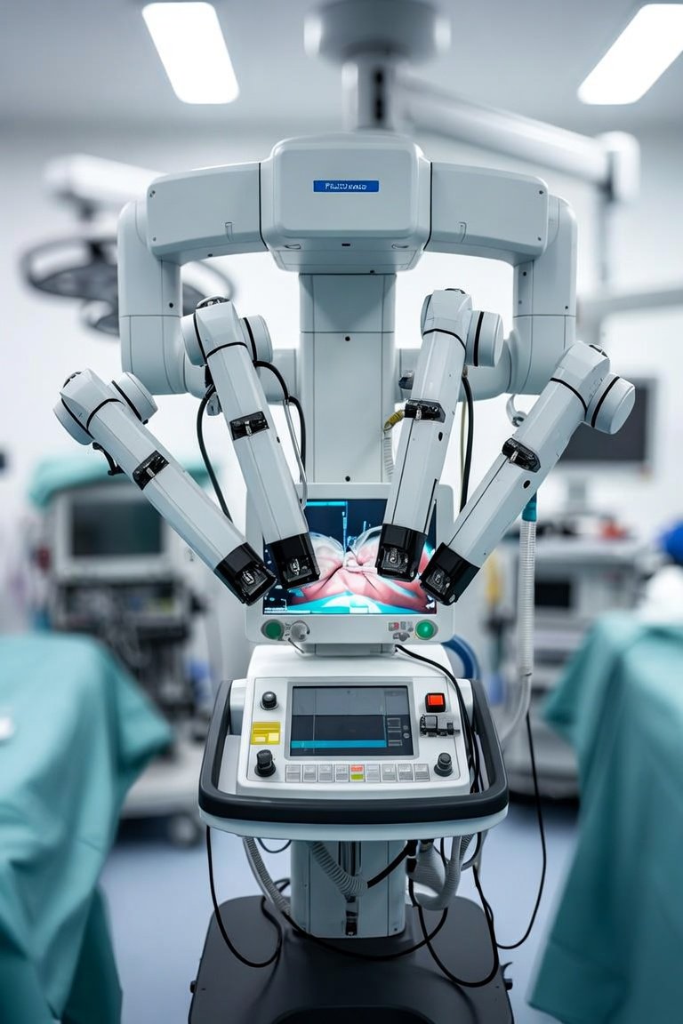
When comparing systems, you have two main categories. First, integrated robotic platforms like the da Vinci Xi with its Firefly fluorescence imaging or the newer Ion endoluminal system. These are closed ecosystems where AI is embedded. Second, modular AI add-ons that work with standard laparoscopes or endoscopes. Examples include Activ Surgical’s AR overlay or Caresyntax’s data analytics. The choice depends on your volume and case mix. For high-volume robotic prostatectomy, an integrated system with AI-driven instrument tracking reduces operative time by roughly 15% in my experience. For general surgeons doing occasional laparoscopic cholecystectomy, a modular overlay that flags cystic artery location is more practical and cost-effective.
What you must look for in any AI-assisted system is validation. Ask three questions. Has the algorithm been trained on diverse patient populations? Many early systems were trained on homogeneous datasets and fail on darker skin tones or unusual anatomy. Is the AI explainable? You need to understand why it highlights a structure, not just trust a black box. And critically, does the system integrate with your existing workflow? If it requires extra steps, like pausing to calibrate, surgeons will abandon it. I have seen excellent technology fail because it added three minutes to setup time.
The real-world benefit I see most often is in reducing cognitive load. In a busy OR, AI can act as a second set of eyes that does not get tired. It can track instruments, monitor vital signs, and even detect early signs of hypotension from insufflation pressure changes. But it is a tool, not a replacement. The best outcomes still come from a skilled surgeon who uses AI to confirm their judgment, not to override it.
My closing advice: start small. Pick one AI feature that addresses a specific pain point in your practice. If you struggle with lymph node mapping, try an AI-enhanced fluorescence system. If you want to reduce port-site hernias, use an AI tool that measures fascial closure tension. Test it on ten cases, then evaluate. The technology is moving fast, but your patients trust your experience. AI is here to augment that experience, not replace it.
Innovations & Tech
AI-Assisted Surgery: Separating Hype from Clinical Reality