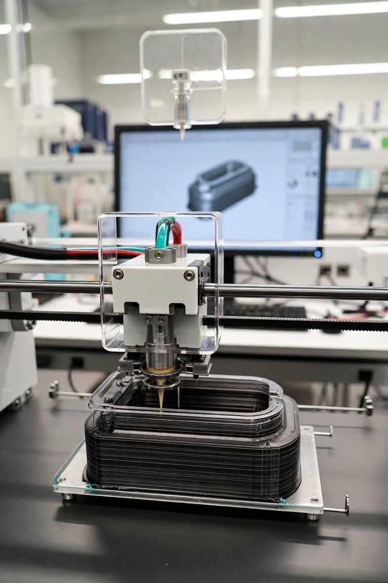
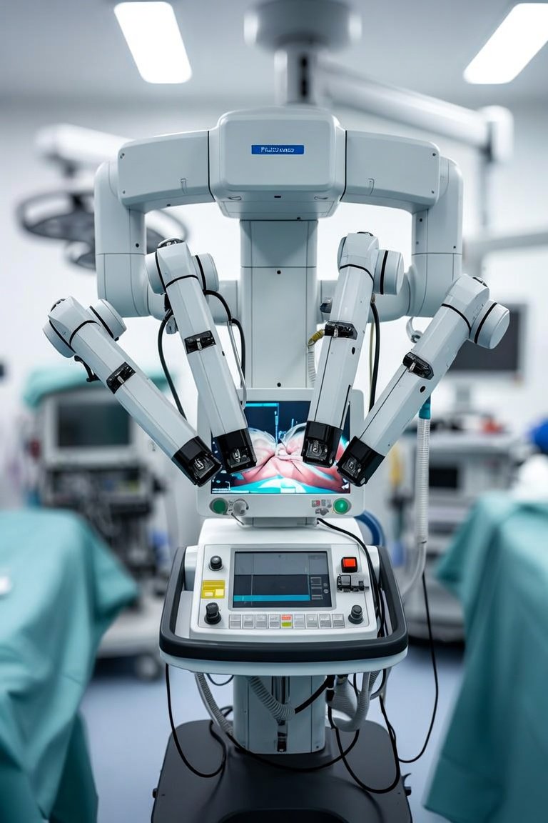
Over the past two decades, I have watched 3D printing evolve from a niche prototyping tool into a transformative force in clinical care. What once seemed like science fiction is now a daily reality in operating rooms and rehabilitation clinics worldwide. I want to share what I have seen work, what has failed, and what you should consider when evaluating these technologies for your facility.
The most profound impact has been in three distinct areas: patient-specific prosthetics, surgical planning models, and biocompatible implants. Let me break down each.
1. Prosthetics and Orthotics. Traditional prosthetic limbs require weeks of casting and fitting. With 3D printing, we can scan a patient’s residual limb in minutes, design a custom socket in software, and print it overnight. The key feature here is MATERIAL CHOICE. For everyday use, I recommend nylon-based filaments like PA12 for their strength and flexibility. For pediatric patients, flexible TPU (thermoplastic polyurethane) offers better comfort and growth accommodation. The cost savings are dramatic: a traditional prosthetic hand can run $5,000 to $10,000, while a 3D-printed version costs under $500 in materials.
2. Surgical Planning and Training Models. This is where 3D printing has saved the most lives in my experience. By converting CT and MRI scans into physical models, surgeons can rehearse complex procedures. For example, in craniofacial reconstruction, a printed skull model allows the team to pre-bend titanium plates and plan osteotomies. I have seen operative times drop by 30 to 40 percent when using these models. The critical specification is RESOLUTION: look for printers capable of 50-micron layer height for fine anatomical detail. Materials like gypsum-based composites offer excellent color differentiation for distinguishing bone, vessels, and tumors.
3. Bioprinting Organs and Tissues. This is the frontier that gets the most headlines, but we must be honest about where we stand. Today, we can print vascularized skin grafts, bone scaffolds, and even functional liver tissue for drug testing. However, whole organ printing for transplantation remains years away. The practical breakthrough has been in SCAFFOLD PRINTING: using hydrogels infused with growth factors to create structures that guide natural tissue regeneration. For bone repair, I have seen excellent results with calcium phosphate-based inks that mimic trabecular bone architecture.
When comparing systems, focus on three factors. First, STERILIZATION COMPATIBILITY: not all printed materials can withstand autoclaving. For surgical tools, I prefer printers using PEEK (polyether ether ketone) filament, which is biocompatible and steam-sterilizable. Second, SOFTWARE INTEROPERABILITY: ensure your printer can accept DICOM files directly from your PACS system. Third, SPEED VERSUS QUALITY: a printer that produces a kidney model in 4 hours at 100-micron resolution may be more practical than one that takes 12 hours for 30-micron detail.
What to look for in a clinical 3D printing system: a closed-loop material system to prevent contamination, FDA-cleared materials for patient contact, and a dedicated medical application specialist for training. Do not buy a printer without seeing it produce a patient-specific model from your own imaging data.
My closing recommendation is straightforward: start with surgical planning models. They require the least regulatory overhead, deliver immediate clinical value, and build the workflow expertise needed for more advanced applications. Once your team masters model generation, move to patient-specific cutting guides, then to permanent implants. The technology is ready. The question is whether your institution is prepared to integrate it into standard of care. I have seen hospitals that hesitated for years, and I have seen early adopters who now cannot imagine practicing without it. The choice is clear.
Innovations & Tech
3D Printing in Medicine: From Bench to Bedside